
Pars plana vitrectomy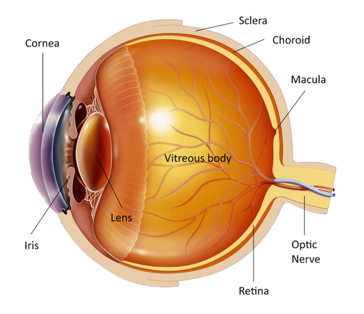
Pars plana vitrectomy is an eye operation where the vitreous gel is removed by small instruments that enter the eye through tiny holes made on its wall. This operation is performed for:
The operation is performed under local or general anesthesia after discussion with your doctor. It may have a long duration (2-3 hours) depending on the condition of your eye and the seriousness of your eye disease. The surgical instruments enter the eye from 3 minuscule holes created on the white of the eye (sclera). The vitreous is removed from the eye and several surgical intraocular maneuvers are made to treat your disease. During the operation the liquid removed from the eye is replaced by a specially manufactured saline so that after the procedure the eye will function normally without the vitreous. At the end of the operation the little holes usually close without any sutures but if necessary small absorbable sutures may be used to close them. The first days after the operation the sutures may cause the feeling of a foreign body in the eye. Usually after the operation the eye is not painful. If a scleral buckle for retinal detachment has been used it may cause some irritation for a few days postoperatively and the eye may appear swollen. You can take painkillers, which will relief your discomfort. And be sure that the doctor does not remove the eye from the socket to operate on it. 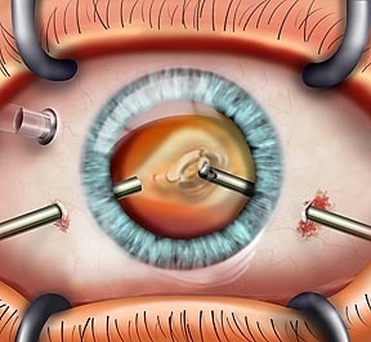
In some cases a gas bubble or silicone oil must be put in your eye in order to keep the retina against the eye wall. For this support to be effective the patient must keep his/her head in the position (posture) advised by his doctor (face down, right or left cheek down) for a period of 7-10 days after the operation, for 50 minutes every hour (10 minutes break). The head position is very important for the success of the operation and should be done reliably. You will be able to eat or go to the toilet without problems. Silicone oil will have to be removed in most cases few months after the original operation with another operation.
You will be able to return home the same or the next day. If a gas bubble is used the vision will be very blurred immediately after the operation, but it will get gradually better within the next 4-8 weeks depending on the type of gas used, as the gas gets absorbed and replaced by fluid. You should not travel by plane while the gas bubble is in your eye. Vitrectomy may rarely cause eye infection or retinal detachment for which 1 or more further operations may be necessary. It may also expedite the development of cataract. Ιf you have any questions about your disease or its treatment do not hesitate to consult you doctor, who will discuss it in details with you. |
The information provided in this web site is not a substitute for professional medical care by a qualified doctor or health care professional. Always check with your doctor if you have concerns about your eye condition or treatment. The authors of this web site are not responsible or liable, directly or indirectly, for any form of damages whatsoever resulting from the information contained in or implied by the information on this site. Information for patients is provided only as a guide.
Copyright Vlassis Grigoropoulos © 2020
Copyright Vlassis Grigoropoulos © 2020


|
Design: Vlassis Grigoropoulos