
Angiography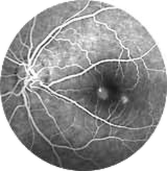
Fluorescein angiography is a very useful examination that gives us valuable information on the blood circulation and the condition of the blood vessels in the retina.
During the examination a dye called fluorescein is injected intravenously and the patient puts his head on a camera like instrument. Within seconds the dye reaches the blood vessels of the eye. Then the camera, fitted with special filters, takes photographs of the retina and blood vessels as the dye fluoresces while is circulating in them. When there is impaired blood circulation, such as swelling, leakage, abnormal blood vessels, then the formations of the dye help us to the disease diagnosis and treatment. Using digital camera provides us with higher quality images and allows for more immediate evaluation of the results. 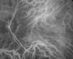
The test is short, you'll see many strong flashes of light and you should not do it if you are allergic to fluorescein or other dyes. For 24 hours after the examination the color of your skin will have a yellow tinge and your urine will be more yellow than usual.
Another angiography that can help us in the diagnosis of many diseases is the indocyanine green angiography. It is done in the same way except for the use of a dye called indocyanine green. It lasts longer than fluorescein angiography (about half an hour) and is used in cases where fluorescein angiography cannot help us, such as bleeding, age-related macular degeneration, a special type of detachments etc. |
The information provided in this web site is not a substitute for professional medical care by a qualified doctor or health care professional. Always check with your doctor if you have concerns about your eye condition or treatment. The authors of this web site are not responsible or liable, directly or indirectly, for any form of damages whatsoever resulting from the information contained in or implied by the information on this site. Information for patients is provided only as a guide.
Copyright Vlassis Grigoropoulos © 2020
Copyright Vlassis Grigoropoulos © 2020


|
Design: Vlassis Grigoropoulos