
Visual fields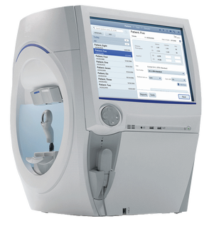
The visual fields test studies not the central vision (how well we can see) but the peripheral sight (how well we perceive the surrounding space). It is used for the diagnosis and follow-up of ophthalmologic diseases such as glaucoma or neurological diseases.
It is a painless examination that requires good concentration. It does not need any preparation or drops in your eyes. Each eye is examined separately with its near glasses. The head is placed in an instrument with a big white dome and while always looking at a central target you should press a switch each time you see a light turning on at the periphery of your vision. The lights may be strong or week, in various points of space and some of them are checking whether you are concentrated. 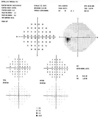
The examination usually is done by an electronic instrument and you should have with you your glasses distant and near. The result is printed out and helps the doctor in the diagnosis and follow-up of your disease. It is important to keep your appointments because the examination must be repeated at regular intervals.
|
The information provided in this web site is not a substitute for professional medical care by a qualified doctor or health care professional. Always check with your doctor if you have concerns about your eye condition or treatment. The authors of this web site are not responsible or liable, directly or indirectly, for any form of damages whatsoever resulting from the information contained in or implied by the information on this site. Information for patients is provided only as a guide.
Copyright Vlassis Grigoropoulos © 2020
Copyright Vlassis Grigoropoulos © 2020


|
Design: Vlassis Grigoropoulos