
Epiretinal membrane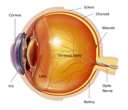
The eye is like a photographic camera. It has the lens and an opening in front that help in the focusing of the objects on the retina. The retina is a thin membrane that is sensitive to the light, as the film in the photographic camera. The macula is found in the center of the retina, where the light focuses. It is responsible for what we see in front of us, for activities such as writing and reading and for the perception of colors. The rest of the retina is responsible for the peripheral vision.
The cavity in front of the retina is full of a transparent jelly like substance that is called vitreous body. In birth and at young age the vitreous is compact and homogeneous but with aging it starts shrinking and becoming liquid and as a result of this it finally gets detached from the retina. This process is called "posterior vitreous detachment". You may have symptoms like floaters but without other consequences. In some cases however, during the posterior vitreous detachment where the vitreous is more adherent to the retina, a small break on the inner surface of the retina may occur. If this happens to the central retina, the macula, a healing process starts with mobilization and displacement of retinal cells, which spread along the retinal surface in an effort to heal the damaged area. This thin membrane of scarring tissue is called "epiretinal membrane" and can lead to mechanical distortion and wrinkling of the retina. 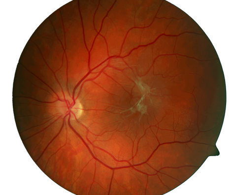
In most cases the healing process is mild and leads to a very thin cellular membrane on the retinal surface. These cells are transparent and do not cause any serious visual symptoms. In some cases however, this process develops into an overproduction of cells that form a thicker, opaque membrane on the retinal surface. Even in these cases the evolution is slow and finally the extension of the membrane is haltered. With time the cells start to contract exerting traction and causing wrinkling of the membrane. If this happens to the macula and because the cells are attached to the retinal surface, the retina itself starts getting wrinkled with onset of visual symptoms.
Central vision may be blurred or distorted and objects get a strange shape and size. This usually happens gradually within months. Because the macula is responsible for vision in details you may have difficulties to read, write and recognize small objects and faces. It is not painful and does not ever lead to blindness because it affects only the central vision. The peripheral vision remains unaffected. This means that all patients with epiretinal membrane have enough vision to maintain their independence and move around with relative comfort. In some cases it may appear in the other eye. 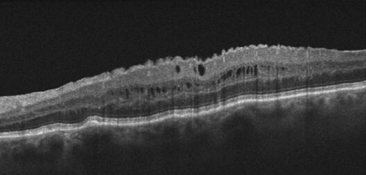
If you think that you are having symptoms of the disease you must visit an ophthalmologist. First he will measure your vision in both eyes. Then drops will be put in your eyes to dilate the pupil and examine the retina. You'll have to wait about half an hour for the drops to work. You will have blurred vision for a few hours and you will be sensitive to the light afterwards and therefore you should not drive home after the examination. Then a special contact lens will be put on your cornea to examine the retina and the macula. Your ophthalmologist may ask you to do a fluorescein angiography (intravenous injection of a dye and pictures of the retina taken with a camera) or an Optical Coherence Tomography test (taking tomographic images of the retina using light) to assess the membrane and to see if there is edema (swelling) or leaking of the membrane in the retina.
In most cases there is no need for any treatment because the symptoms are minimal. However, patients that have significant trouble to see and to do their daily activities, such as reading or driving, or are having findings on fluorescein angiography are candidates for treatment. The treatment is a special surgical procedure called "vitrectomy". This procedure is done under local or general anesthetic after discussion with your doctor and lasts approximately 1-1.5 hours. The surgical instruments enter the eye from 3 minuscule holes created on the white of the eye (sclera). The vitreous is removed from the eye and mainly from the macula relaxing any traction exerted on the retina. Then special microsurgical instruments are used to remove the membrane from the retina. During the operation the liquid removed from the eye is replaced by a specially manufactured saline so that after the procedure the eye will function normally without the vitreous. At the end of the operation the little holes close without any sutures. You will be able to return home the same or the next day. In few cases a gas bubble may be put in your eye and you will have to posture your head (keep it to a particular position) for 7-10 days. You should not travel by plane while the gas bubble is in your eye. And be sure that the doctor does not remove the eye from the socket to operate on it. In most cases after the operation the distortion goes away and the vision improves but the degree of improvement varies in each individual. Vitrectomy may rarely cause retinal detachment for which 1 or more operations may be necessary. It may also expedite the formation of cataract. If you have any questions about your disease or its treatment do not hesitate to consult you doctor, who will discuss it in details with you. |
The information provided in this web site is not a substitute for professional medical care by a qualified doctor or health care professional. Always check with your doctor if you have concerns about your eye condition or treatment. The authors of this web site are not responsible or liable, directly or indirectly, for any form of damages whatsoever resulting from the information contained in or implied by the information on this site. Information for patients is provided only as a guide.
Copyright Vlassis Grigoropoulos © 2020
Copyright Vlassis Grigoropoulos © 2020


|
Design: Vlassis Grigoropoulos