
Retinal detachment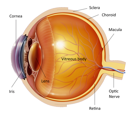
The eye is like a photographic camera and the retina is the film. The retina is a thin membrane of nerve tissue that covers the inner surface of the eye. The light enters the eye through the cornea and the lens and focuses on the retina. Then the image is formed and is sent through the optic nerve to the brain for processing. It is similar to the photographic film that must be developed in order to have the pictures.
Usually the retina is attached to the inner surface of the eye. If a hole is formed on the retina then fluid may pass under the retina, weaken its connections and detach the retina - as the wallpaper comes off the wall. When this happens the retina cannot form a clear image and the vision becomes weak and blurry. 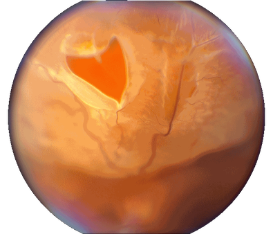
The retinal detachment is a rare condition that affects approximately 1 in 10000 persons. It happens more often to people of middle or old age with myopia. Conditions like diabetes, cataract operation or trauma may cause retinal detachment. Rarely young individuals may have weak areas on the retina.
The most common symptom is a shadow that spreads in front of the eye. You may also have flashes - like the flask of a camera - or many sudden small dots of various shapes in front of your eye. Those symptoms are never painful. If you have them you will have to visit an ophthalmologist as soon as possible. Prompt treatment will minimize the damage on your vision. If you visit an ophthalmologist early enough then laser or freezing of the retinal hole may be sufficient. This treatment is usually done under local anesthetic. Often however an operation is necessary to treat the retinal hole. It is performed under local or general anesthetic and is successful in 80-90% of cases. 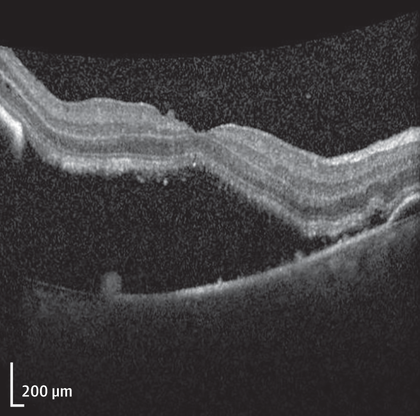
There are 2 different operations for a retinal detachment. One, called scleral buckle, has a piece of plastic sutured to the wall of the eye at the area of the retinal hole, pressing the retina from the outside. This plastic usually remains on the eye but if needed it may be removed with another operation. It may also cause myopia.
In the other operation, called pars plana vitrectomy, the vitreous that fills the eye is removed through instruments that enter the eye through tiny holes and is replaced by air, gas or silicone oil, pressing the retina from inside. You will have to keep your head in the position (posture) advised by your doctor (face down, right or left cheek down) for a period of 7-10 days after the operation, for 50 minutes every hour (10 minutes break). You should not travel by plane while the gas bubble is in your eye. This operation may cause cataract. If silicone oil is used it will have to be removed in most cases few months after the original operation with another operation. If a gas bubble is used the vision will be very blurred immediately after the operation, but it will get gradually better within the next 4-8 weeks depending on the type of gas used, as the gas gets absorbed and replaced by fluid. The choice of the type of operation that is suitable for you will be done by your doctor. The operation usually is not very painful but your eye may be swollen and bothersome for a few days after. And be sure that the doctor does not remove the eye from the socket to operate on it. The vision that you will have after a successful operation depends on the extent and duration of retinal detachment. The shadow caused by the retinal detachment will disappear in all cases after a successful operation. But if the detachment affects the central vision then the central vision may not improve considerably. The longer the central part of the retina remains detached the less likely is that the central vision will be restored. But even in that case you will retain useful peripheral vision. Usually you will be able to go home the next day after the operation. You may resume your daily activities, including sex, as soon as you want, taking of course all precautions that your doctor will advise you. If you are not operated or if the operation fails then you will loose all your site in this eye. In case of failure of the first operation you may need 1 or more further operations to treat the retinal detachment. If you have a family history of retinal detachment or your doctor finds weak areas on your retina he may advise you to have laser or freezing of the eye to prevent the development of a retinal detachment. Unfortunately, in most cases retinal detachment cannot be prevented. You will not develop a retinal detachment if you tire your eyes or if you bend over or lift heavy things. If you get a retinal detachment in one eye then you have increased chances (10%) to develop a retinal detachment in the other eye. Ιf you have any questions about your disease or its treatment do not hesitate to consult you doctor, who will discuss it in details with you. |
The information provided in this web site is not a substitute for professional medical care by a qualified doctor or health care professional. Always check with your doctor if you have concerns about your eye condition or treatment. The authors of this web site are not responsible or liable, directly or indirectly, for any form of damages whatsoever resulting from the information contained in or implied by the information on this site. Information for patients is provided only as a guide.
Copyright Vlassis Grigoropoulos © 2020
Copyright Vlassis Grigoropoulos © 2020


|
Design: Vlassis Grigoropoulos