
Intraocular pressure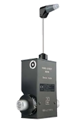
The intraocular pressure measurement is a test that checks the pressure of the eye, the intraocular pressure. It measures the pressure of the fluid inside the eyes, to see if it is within normal limits. The operation is based on the resistance encountered by the flattening of a small area of the cornea.
The examination is done by instilling local anesthetic and a dye, which fluoresces when stimulated by blue light, in both corneas. Then the tonometer touches the cornea of each eye under blue illumination. As the patient sits on the slit lamp, he sees a small circle of intense light approaching his eye. The examination is quick and completely painless with no effect on the vision. 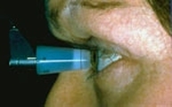
The high pressure inside the eye can cause irreversible damage to the optic nerve, causing what is known as glaucoma, which needs treatment with drops. People suffering from glaucoma should regularly check the intraocular pressure to determine whether treatment is effective or needs amendment.
|
The information provided in this web site is not a substitute for professional medical care by a qualified doctor or health care professional. Always check with your doctor if you have concerns about your eye condition or treatment. The authors of this web site are not responsible or liable, directly or indirectly, for any form of damages whatsoever resulting from the information contained in or implied by the information on this site. Information for patients is provided only as a guide.
Copyright Vlassis Grigoropoulos © 2020
Copyright Vlassis Grigoropoulos © 2020


|
Design: Vlassis Grigoropoulos