
Central retinal vein occlusion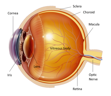
The eye is like a photographic camera. It has the lens and an opening in front that help in the focusing of the objects on the retina. The retina is a thin membrane that is sensitive to the light, as the film in the photographic camera. The macula is found in the center of the retina, where the light focuses. It is responsible for what we see in front of us, for activities such as writing and reading and for the perception of colors. The rest of the retina is responsible for the peripheral vision.
The retina is nourished by the blood flow, which provides the nutrients and the oxygen that the nerve cells need for proper operation. When there is a blockage in the veins of the retina then we have what we call a retinal vein occlusion. Central retinal vein occlusion is the blockage of the main vein of the retina. (When there is blockage of the small veins of the retina it is called Branch retinal vein occlusion). The occlusion of the vein causes leakage of blood and liquid from the blood vessels through their wall into the retina. When the main retinal vein is closed the nerve cells of the entire retina may suffer significant losses. Because the macula, the part of the retina responsible for central vision, is affected by the occluded veins part of central vision may be lost. There are 2 types of Central retinal vein occlusion:
The most common symptom of Central retinal vein occlusion is loss of vision or blurring of part or all of the vision in one eye. It is painless and can occur suddenly or gradually over a period of several hours or days. Sometimes there is a sudden and complete loss of vision. Another symptom may be the so called floaters. Because the retinal blood vessels are not functioning properly new blood vessels are formed in the eye, as nature is trying to correct the problem and supply the eye with blood. Unfortunately, these blood vessels are of bad quality, feeble and are formed in the wrong part of the eye - on the retinal surface and inside the vitreous cavity. As a consequence, they bleed easily and form the floaters that we may see in front of our eye. In some serious cases of Central retinal vein occlusion a painful eye with high intraocular pressure may develop called "neovascular glaucoma". Neovascular glaucoma appears in the ischemic type of Central retinal vein occlusion and is caused by the abnormal blood vessels that grow inside the eye and increase its pressure. It is a serious complication that leads to severe pain and extended visual loss. It usually takes more than 3 months for the neovascular glaucome to develop. It is associated with aging and usually occurs in people aged 50 years and older. Hypertension, glaucoma and atherosclerosis are usually associated with it. People with diabetes are at increased risk for Central retinal vein occlusion. If you have sudden vision loss you should visit an ophthalmologist. First he will measure the vision in both eyes, he will examine the pupillary reflex, he will measure the intraocular pressure and will perhaps examine your visual fields. Then he will put drops in both your eyes to dilate the pupils and examine the retina. You will have to wait for about half an hour for the drops to work. You will have blurred vision for a few hours and you will be sensitive to the light afterwards and therefore you should not drive home after the examination. Then a special contact lens will be put on your cornea to examine the retina and the macula. Your ophthalmologist will ask you to do a fluorescein angiography (intravenous injection of a dye and pictures of the retina taken with a camera) and an Optical Coherence Tomography test (taking tomographic images of the retina using light) to assess the macula and to see if there is edema (swelling) or leaking of the retina due to the vein occlusion or abnormal blood vessels. In addition, you will need tests to determine the levels of sugar and cholesterol in your blood. People under 40 should be checked for blood clotting problems. 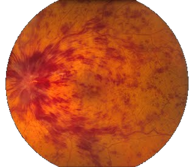
Because the clot in the vein cannot be removed there is no cure for Central retinal vein occlusion. Some people regain some degree of vision but full visual recovery is a rare event.
Discovering what causes the occlusion is the first step in treatment. Your ophthalmologist may recommend a waiting and observation period after diagnosis. During the course of the disease, many patients will experience swelling in the central region of the macula. This swelling, called macular edema, can last for more than a year. The treatment for macular edema is laser. Laser is a light beam that can be focused on the retina with high precision. Thus we can close the leaking retinal blood vessels. Laser is done in the clinic and is usually painless. After you have drops put in your eyes to dilate the pupil you sit on the slit lamp, a special contact lens is put on the cornea to help focus the laser on the retina and you will see many strong flashes of light. The treatment for the leaking blood vessels doesn't last long and its main objective is to stabilize vision by closure of the leaking blood vessels that prevent the macula from functioning properly. The treatment for the new blood vessels however lasts longer and sometimes can be painful. If this is the case then a local anesthetic injection can be done around your eye to relieve you. The side effects of laser for the leaking blood vessels may be a small reduction in visual acuity. The side effects of laser for the new blood vessels may be more prominent. Part of your peripheral vision is often lost. Your night vision and the perception of colors may also be affected. It is very important however to know that there is no treatment without side effects and that the dangers from no treatment are far greater than those from laser itself. It has been found that injection of a substance (eg Lucentis etc) into the eye that inhibits the abnormal blood vessels and reduces leakage is able to stop the progression of the disease and improve vision. These injections are performed in theatre painlessly under sterile conditions and they must often be repeated at intervals of 1-2 months. A small device may also be injected into the eye releasing slowly over several months cortisone into the eye and thereby reducing the swelling of the retina. If you have any questions about your disease or its treatment do not hesitate to consult your doctor, who will discuss it with you in detail. |
The information provided in this web site is not a substitute for professional medical care by a qualified doctor or health care professional. Always check with your doctor if you have concerns about your eye condition or treatment. The authors of this web site are not responsible or liable, directly or indirectly, for any form of damages whatsoever resulting from the information contained in or implied by the information on this site. Information for patients is provided only as a guide.
Copyright Vlassis Grigoropoulos © 2020
Copyright Vlassis Grigoropoulos © 2020


|
Design: Vlassis Grigoropoulos