
Glaucoma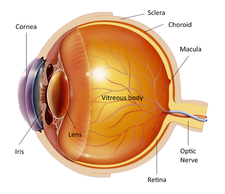
Glaucoma is a group of diseases that cause damage to the optic nerve head as it comes out of the eye. The optic nerve transmits information from the retina, the light sensitive membrane of the eye, to the brain where the image is processed.
The eye must have a specific internal pressure in order to function normally. In some people the damage is due to elevated intraocular pressure. In other people, despite the normal intraocular pressure, the damage occurs from a weakness of the optic nerve. In most cases both factors coexist. The intraocular pressure is independent from the blood pressure. The cells behind the iris of the eye are producing a liquid called aqueous. Aqueous passes through an opening in the middle of the iris called pupil to the front of the eye. It gets out of the eye through minuscule channels that exist at the angle formed by the cornea and the iris in order to get back into the blood circulation. Under normal conditions the same amount of aqueous that is produced gets absorbed into the blood stream but if there is a problem with absorption or if more aqueous is produced then we have an increase of intraocular pressure. Aqueous has nothing to do with tears. High intraocular pressure damages the optic nerve. The extent of the damage depends on the level of the intraocular pressure, its duration and if there is reduced blood supply or weakness of the optic nerve. Very high and sudden elevation of the intraocular pressure will cause an immediate damage to the optic nerve. A smaller intraocular pressure increase will damage the optic nerve at a slower rate and vision will gradually deteriorate if not treated. There are 4 main types of glaucoma:
The factors that increase the danger of developing glaucoma are:
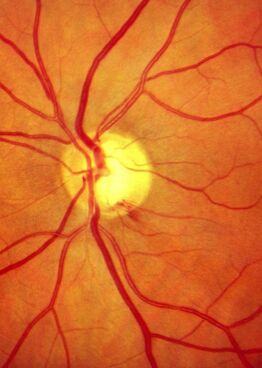
The danger in open angle glaucoma is that your eye seems absolutely healthy. There is no pain and the vision appears normal although it has already started to deteriorate. The initial visual fields defect has the shape of an arc located a little above or below the central vision. If left untreated this defect will gradually spread around until only the central vision remains intact, called "tunnel vision", with total vision loss at the last stages of the disease.
Since glaucoma appears more often after the age of 40 you should check your intraocular pressure every 1-2 years as well as your optic nerve under the microscope and the visual fields. Open angle glaucoma is treated by lowering the intraocular pressure. This is initially achieved by eye drops that reduce the production of aqueous or increase its outflow. If this proves ineffective then surgery will be necessary. Although any loss of vision is permanent, early diagnosis and prompt treatment can minimize the damage and maintain good visual acuity. In acute or closed angle glaucoma the intraocular pressure increases suddenly due to narrow anterior chamber angle that does not allow the aqueous to leave the eye. The sudden raise in intraocular pressure is very painful. The eye is red, the vision is blurred, there is nausea and vomiting. Sometimes you may have mild attacks of high intraocular pressure, especially at night, with blurred vision, color rings around the lights and some degree of discomfort. In the acute phase you should go to the hospital immediately to treat the pain and the high intraocular pressure with medication. This is usually achieved within a few hours and the vision is restored. Then your doctor will advise you to make a small hole on the iris of both eyes with laser to avoid another similar attack in the future. The operation is not painful. If acute glaucoma is left untreated then vision can be seriously and permanently damaged. You can drive if the visual fields defect is not advanced. If you have any questions about your disease or its treatment do not hesitate to consult you doctor, who will discuss it in details with you. |
The information provided in this web site is not a substitute for professional medical care by a qualified doctor or health care professional. Always check with your doctor if you have concerns about your eye condition or treatment. The authors of this web site are not responsible or liable, directly or indirectly, for any form of damages whatsoever resulting from the information contained in or implied by the information on this site. Information for patients is provided only as a guide.
Copyright Vlassis Grigoropoulos © 2020
Copyright Vlassis Grigoropoulos © 2020


|
Design: Vlassis Grigoropoulos