
Age related macular degeneration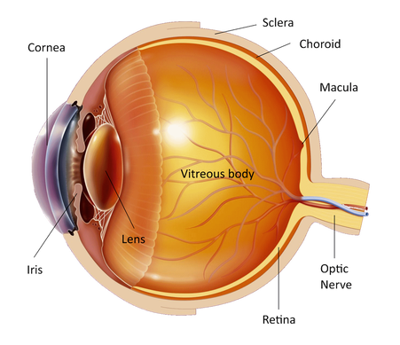
The eye is like a photographic camera. It has the lens and an opening in front that help in the focusing of the objects on the retina. The retina is a thin membrane that is sensitive to the light, as the film in the photographic camera. The macula is found in the center of the retina, where the light focuses. It is responsible for what we see in front of us, for activities such as writing and reading and for the perception of colors.
Sometimes the cells of the macula suffer damage and they stop working. We do not know why but it usually happens as the person gets older. It is called age related macular degeneration. Because it is usually related with age it presents in both eyes but mostly at different times. In certain eyes the cells simply stop working, like the colors get washed out in old photographs. This type of degeneration is called "dry". In other eyes a scar is formed in the macula because of leakage from the blood vessels and it is called "wet" degeneration. 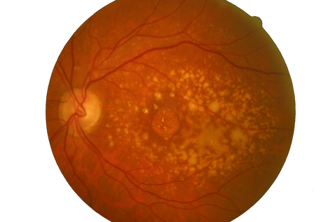
Children and young individuals can also present macular degeneration, which is hereditary and is called “macular dystrophy”. Often many members of a family present the same disease and if this happens in your family then you will need to check your eyes regularly by an ophthalmologist.
Macular degeneration is the most common cause of low vision in individuals over 60. It is not painful and it does not lead to blindness because it only affects the central vision. The peripheral vision remains unaffected. This means that all patients with macular degeneration have enough vision to maintain their independence and move around with relative comfort. 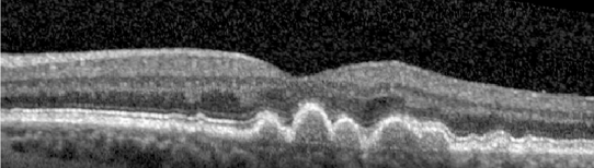
In the early stages the central vision may be blurry or deformed and the objects have curious shape and size. This can happen fast or progressively within months. You may be sensitive to light or you may see lights that don't exist and this can cause some nuisance but no pain.
The macula is the part of the eye that allows you to see in detail and at the advanced stages of the disease you will often have a dark spot in the middle of your central vision. This makes activities such as reading, writing and recognition of small objects and faces very difficult. 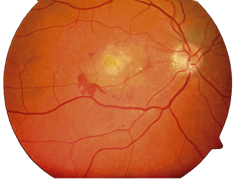
If you think that you have symptoms of the disease or you already have the disease in one eye and you are afraid that the other eye is affected then you will have to visit your ophthalmologist promptly. First he will measure your vision in both eyes. Then he will put drops in both your eyes to dilate the pupils and examine the retina. You will have to wait for about half an hour for the drops to work. You will have blurred vision for a few hours and you will be sensitive to the light afterwards and therefore you should not drive home after the examination.
Your ophthalmologist will ask you to do a fluorescein angiography to see what exactly is happening to your macula and decide for the best treatment options for you. During this test a dye will be injected intravenously into your arm, which will reach your eyes within seconds. It is not painful but you may feel a slight nausea. Then a series of quick photos of your retina will be taken within minutes. After the test you will notice that your skin and urine have a yellow color but this will go away soon. Side effects are very rare with the main one being an allergic reaction, which will need immediate attention. 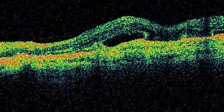
Another test that is necessary for the diagnosis and the follow-up of the disease is the Optical Coherence Tomography (OCT). It is a painless and quick examination of the retina and macula that shows, like a kind of computerized tomography but without radiation, all the layers of the retina and the changes of the disease.
The wet type of age related macular degeneration, if diagnosed early, can be treated with laser. This can be done at the clinic and is painless. You sit as the slit lamp and a special lens is put on your eye to focus the laser on the retina. The treatment does not last long and you will see many flashes of light. Unfortunately, in most cases the damage is at the center of the macula and conventional laser can not be used because the scar formed will deteriorate the central vision. Lately, the injection of a drug (eg Lucentis etc) that causes regression of the abnormal blood vessels, into the eye can not only stop the course of the disease but also improve the vision. These injections are done in theater painlessly, under sterile conditions and often may need to be repeated every 1-2 months. Another treatment that is used in some cases is called "photodynamic" therapy. A special drug is injected intravenously and after few minutes a special lens is put on your eye to focus the laser on the retina. Then a special laser is applied that activates the injected drug and stops the leaking of the blood vessels. After the treatment you will be given special glasses and you will have to avoid the sunlight for 2 days. You should follow the advice of your doctor. Often, this treatment may need to be applied more than once every few months until the leaking of the blood vessels stops permanently. This treatment is done mainly to stabilize the vision and prevent it from worsening. No treatment is currently available for the dry type of age related macular degeneration. There are a lot of low vision aid instruments that you can use, from magnifying lenses to closed circuit television sets. Many research projects are currently under way for the investigation of the causes and treatment possibilities of the disease. If you have any questions about your disease or its treatment do not hesitate to consult you doctor, who will discuss it in details with you. |
The information provided in this web site is not a substitute for professional medical care by a qualified doctor or health care professional. Always check with your doctor if you have concerns about your eye condition or treatment. The authors of this web site are not responsible or liable, directly or indirectly, for any form of damages whatsoever resulting from the information contained in or implied by the information on this site. Information for patients is provided only as a guide.
Copyright Vlassis Grigoropoulos © 2020
Copyright Vlassis Grigoropoulos © 2020


|
Design: Vlassis Grigoropoulos