Vlassis Grigoropoulos MD PHD
  • Home
    • Curriculum Vitae
    • Private practice
  • Tests
    • Angiography
    • Corneal pachymetry
    • Fundoscopy
    • Intraocular pressure
    • Optical Coherence Tomography
    • Slit lamp
    • Visual fields
  • Diseases
    • Branch retinal vein occlusion
    • Cataract
    • Central retinal vein occlusion
    • Diabetic retinopathy
    • Epiretinal membrane
    • Glaucoma
    • Macular degeneration
    • Macular hole
    • Posterior vitreous detachment
    • Retinal detachment
    • Vitrectomy
  • Activities
    • TVRS 2021
    • Dunant 2019
    • TVRS 2017
    • OMMA 2016
    • QuORe 2016
    • TVRS 2016
    • Diabetic screening
    • Ocriplasmin
  • GR

Slit lamp

Picture
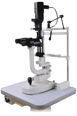
The slit lamp is basically a microscope. It has a light source with variable beam width and intensity with which the ophthalmologist can examine your eyes in magnification. 

This instrument is mainly used to check the front part of the eye such as the cornea, the iris and the lens. 

Using special lenses we can also look into the vitreous and the back of the eye, the retina. 

The ophthalmologist sits on one side of the slit lamp and the patient on the other. It is possible to use special lenses that are placed in front or onto the eye in order to examine the posterior segment of the eye such as the vitreous, the optic nerve and the retina.


The information provided in this web site is not a substitute for professional medical care by a qualified doctor or health care professional. Always check with your doctor if you have concerns about your eye condition or treatment. The authors of this web site are not responsible or liable, directly or indirectly, for any form of damages whatsoever resulting from the information contained in or implied by the information on this site. Information for patients is provided only as a guide.

Copyright Vlassis Grigoropoulos © 2020
Search    Facebook    Linkedin    Youtube    Youtube    Email
Design: Vlassis Grigoropoulos
Proudly powered by Weebly
  • Home
    • Curriculum Vitae
    • Private practice
  • Tests
    • Angiography
    • Corneal pachymetry
    • Fundoscopy
    • Intraocular pressure
    • Optical Coherence Tomography
    • Slit lamp
    • Visual fields
  • Diseases
    • Branch retinal vein occlusion
    • Cataract
    • Central retinal vein occlusion
    • Diabetic retinopathy
    • Epiretinal membrane
    • Glaucoma
    • Macular degeneration
    • Macular hole
    • Posterior vitreous detachment
    • Retinal detachment
    • Vitrectomy
  • Activities
    • TVRS 2021
    • Dunant 2019
    • TVRS 2017
    • OMMA 2016
    • QuORe 2016
    • TVRS 2016
    • Diabetic screening
    • Ocriplasmin
  • GR