
Corneal pachymetry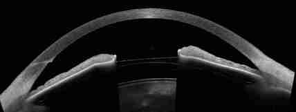
Corneal pachymetry is the measurement of the thickness of the cornea. From recent studies it was found that the thickness of the may be related with the possibility to develop glaucoma.
The applanation tonometer we use for the measurement of the intraocular pressure has been manufactured for a certain base corneal thickness while in the general population corneal thickness normally varies. Therefore, if the thickness that we measure is higher than the base thickness then the real intraocular pressure is lower while if the thickness that we measure is lower than the base thickness then the real intraocular pressure is higher. This is why now days corneal thickness measurement constitutes a routine examination in the investigation of glaucoma. The examination is very short and painless. We put local anesthetic in the eyes and with a special prism we touch the cornea while the instrument is recording its thickness. Then with a special formula we calculate the adjustment of the real intraocular pressure based on our measurements. The examination needs to be done only once unless you are submitted to operations that change the shape of your cornea, such as correction of myopia etc. |
The information provided in this web site is not a substitute for professional medical care by a qualified doctor or health care professional. Always check with your doctor if you have concerns about your eye condition or treatment. The authors of this web site are not responsible or liable, directly or indirectly, for any form of damages whatsoever resulting from the information contained in or implied by the information on this site. Information for patients is provided only as a guide.
Copyright Vlassis Grigoropoulos © 2020
Copyright Vlassis Grigoropoulos © 2020


|
Design: Vlassis Grigoropoulos