
Macular hole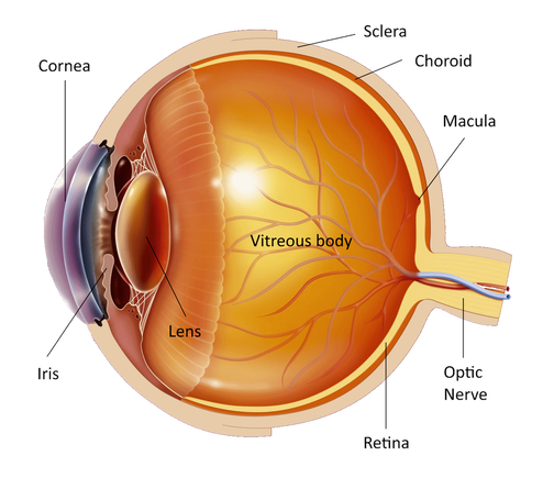
The eye is like a photographic camera. It has the lens and an opening in front that help in the focusing of the objects on the retina. The retina is a thin membrane that is sensitive to the light, as the film in the photographic camera. The macula is found in the center of the retina, where the light focuses. It is responsible for what we see in front of us, for activities such as writing and reading and for the perception of colors. The rest of the retina is responsible for the peripheral vision.
The cavity in front of the retina is full of a transparent jelly like substance that is called vitreous body. At birth and at young age the vitreous has a compact and homogeneous structure but with age it starts shrinking and liquefying and eventually it gets detached from the retina. This process is called "posterior vitreous detachment". There may be symptoms like floaters but without further consequences. In some people however during posterior vitreous detachment a hole on the retina may form to areas where the vitreous is strongly adherent to the retina or to areas where the retina is weaker. If this happens to the macula we then have what we call a "macular hole". Liquefied vitreous may then pass through the hole under the retina and cause a localized retinal detachment. 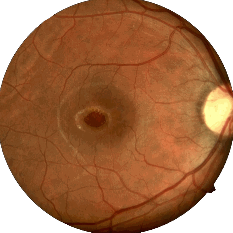
At the early stages of the disease central vision may be blurred or distorted and objects can have a strange shape and size. This usually happens gradually within months. The macula is the part of the eye that allows you to see in detail and at the advanced stages of the disease you will have a dark spot at the center of your vision. This makes activities such as reading, writing and recognition of small objects and faces very difficult. In some cases at the early stages the disease process may revert and the vision may improve. It is not painful and never leads to blindness because it interferes only with the central vision. The peripheral vision remains unaffected. This means that all patients with macular hole have enough vision to maintain their independence and move around with relative comfort. Rarely it may appear in the other eye.
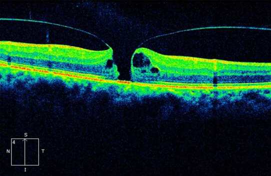
If you think that you are having symptoms of the disease you must visit an ophthalmologist. First he will measure your vision in both eyes. Then drops will be put in your eyes to dilate the pupil and examine the retina. You'll have to wait about half an hour for the drops to work. You will have blurred vision for a few hours and you will be sensitive to the light afterwards and therefore you should not drive home after the examination. Then a special contact lens will be put on your cornea to examine the retina and the macula. Your ophthalmologist may ask you to do a fluorescein angiography (intravenous injection of a dye and pictures of the retina taken with a camera) or an Optical Coherence Tomography test (taking tomographic images of the retina using light) to better assess the hole and its causes (trauma, high myopia etc).
The only treatment of macular hole is a special surgical procedure called "vitrectomy" that can improve your vision. This procedure is done under local or general anesthetic after discussion with your doctor and lasts approximately 1-1.5 hours. The surgical instruments enter the eye from 3 minuscule holes created on the white of the eye (sclera). The vitreous is removed from the eye and mainly from the macula relaxing any traction exerted on the retina. During the operation the liquid removed from the eye is replaced by a specially manufactured saline so that after the procedure the eye will function normally without the vitreous. At the end of the operation a gas bubble will be put in your eye to press on the retina and help the closure of the hole and the little scleral holes close without any sutures. You will be able to return home the same or the next day. For the operation and the gas bubble to be effective you will have to posture your head (keep it to a particular position) face down for 7-10 days after surgery, 50 minutes every hour (10 minutes break). The position of your head is very important for the success of the operation and you should be very consistent. You will be able to eat and go to the toilet without any problem. The vision will be very blurred immediately after the surgery and will gradually improve, within 4-8 weeks depending on the type of gas used, as the gas bubble gets absorbed, until it gets totally replaced by liquid. You should not travel by plane while the gas bubble is in your eye. And be sure that the doctor does not remove the eye from the socket to operate on it. In up to 90% of cases the hole closes and the vision improves but the improvement varies with each patient. In cases where the hole does not close the surgery may be repeated with significant chances of success. A small number of patients may present years after surgery with reopening of the hole and will be offered treatment with re-operation. Vitrectomy may rarely cause retinal detachment for which 1 or more operations may be necessary. It may also expedite the formation of cataract. Ιf you have any questions about your disease or its treatment do not hesitate to consult you doctor, who will discuss it in details with you. |
The information provided in this web site is not a substitute for professional medical care by a qualified doctor or health care professional. Always check with your doctor if you have concerns about your eye condition or treatment. The authors of this web site are not responsible or liable, directly or indirectly, for any form of damages whatsoever resulting from the information contained in or implied by the information on this site. Information for patients is provided only as a guide.
Copyright Vlassis Grigoropoulos © 2020
Copyright Vlassis Grigoropoulos © 2020


|
Design: Vlassis Grigoropoulos